166a Nano-Sized Bioactive Glass In a Biodegradable Polymer: How Advantageous Is Nano-Size?
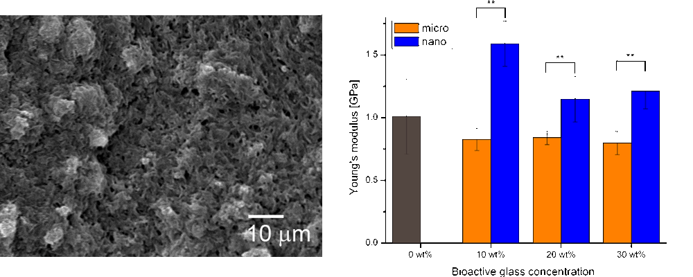
The addition of NBG particles induced a nano-structured topography (Figure) on the surface of the composites not visible on micron-sized particles containing composites. This surface effect observed for NBG composites considerably increased the protein adsorption and had a reinforcement effect on the composite (Figure). Immersion in SBF revealed a high level of in vitro bioactivity for P(3HB)/NBG composites. Proliferation of MG-63 osteoblast-like cells on the various composites demonstrated a good cytocompatibility of all composite materials.
This study revealed that such nanoparticles are a most interesting bioactive filler material for biodegradable polymers in order to prepare advanced composites for tissue engineering.
Figure: Scanning electron microscopy images of a planar section of P(3HB) with 30 wt% nano-sized bioactive glass particles (left). Modulus comparison for various concentrations of micron- and nano-sized bioactive glass particles in P(3HB) composites. **p < 0.01
References
[1] K. Rezwan, Q.Z. Chen, J.J. Blaker and A.R. Boccaccini, Biomaterials, 2006, 27, 3413-31.
[2] S. Loher, V. Reboul, T.J. Brunner, M. Simonet, C. Dora, P. Neuenschwander and W.J. Stark, Nanotechnology, 2006, 17, 2054-61.
[3] T.J. Brunner, R.N. Grass and W.J. Stark, Chem. Commun., 2006, 13, 1384-6.
[4] S.K. Misra, D. Mohn, T.J. Brunner, W.J. Stark, S.E. Philip, I. Roy, V. Salih, J.C. Knowles and A.R. Boccaccini, Biomaterials, 2008, 29, 1750-61.
